
Help Document
[Document] Scanning Electron Microscope Resolution Detection and Acceptance
Release Time:2018-08-01
Scanning electron microscope manufacturers usually provide a blank test report, clearly specifying the test items, indicators, and methods. Testing can be conducted step-by-step, from easy to difficult. Some manufacturers also provide the instrument's factory inspection report for reference. Users compare the measured results with the technical indicators; if they meet the requirements, mark "OK" in the corresponding item column. After completing the item-by-item verification, if the user has no objections, both parties sign the test report. The duration of the electron microscope debugging and testing depends on the quality of the electron microscope itself and the technical level of the manufacturer's engineers. In addition, the commissioning and acceptance of field emission microscopes are much more complex than conventional microscopes. Electron gun baking alone takes at least 2-3 days. If the technical indicators can be tested within a week, it is considered very smooth.
Scanning electron microscopes have multiple performance and technical indicators, such as: resolution, magnification error, low-magnification distortion, beam current stability, sample contamination rate, vacuum degree, sample exchange vacuum time, start-up to working vacuum time, sample stage displacement distance, etc., all of which have quantitative indicators. Here, the focus is on resolution.
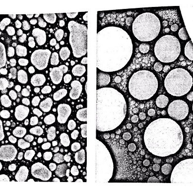
Resolution is an important indicator of an electron microscope; it reflects the overall performance of the instrument. Different types of electron microscopes have different resolutions, indicating differences in the scale at which the instrument enters the microscopic field. This is also a selling point of different grades of electron microscopes. Users are very concerned about whether this indicator meets the requirements. For the manufacturer's engineers, taking resolution photos is also a technical challenge. They need to adjust all aspects of the electron microscope to the optimal state, select almost extreme operating conditions, take repeated photos until they produce satisfactory photos. The upper left image shows a high-resolution photo of gold particles on carbon, with a magnification of 300,000 times, an accelerating voltage of 5 kV, a working distance of 5 mm, and a beam spot selection of 1, which is the smallest size.
1. Resolution Standard Sample
The standard sample is gold particles on a carbon substrate. The advantages of this standard sample are as follows: the atomic numbers of carbon and gold differ greatly, the standard sample has high natural contrast, making it easy for the electron microscope to select the smallest beam spot for high-resolution imaging; the standard sample has good electrical and thermal conductivity, which is stable for the electron beam; the gold particle size is 2 nm to 30 nm, suitable for images with magnifications of 50,000 times or more. This standard sample has been adopted by various electron microscope companies.
Another standard sample is tin balls on a carbon substrate. The sample has high natural contrast; it is more resistant to electron beam irradiation than gold particles. When magnified at high magnification, repeated astigmatism correction and focusing at a certain point will not cause the tin ball to become contaminated and black; the tin ball size is 3 nm to 30 μm, this wide size range is suitable for instruments with magnifications ranging from low to high. The spherical shape facilitates astigmatism correction.
Usually, both standard samples can be placed in the electron microscope simultaneously. First, use the tin balls to adjust the instrument, and then use the gold particles to take resolution photos. The upper right image shows a high-magnification photo of tin balls, with a magnification of 100,000 times, an accelerating voltage of 15 kV, a working distance of 5 mm, and a beam spot selection of 3. Many tin balls of different diameters are visible.
2. Resolution Evaluation
The resolution photo is a secondary electron image of gold particles. The shooting conditions vary slightly among companies. For field emission electron microscopes, the magnification is selected at 200,000 times or 300,000 times; the accelerating voltage is 15 kV to 30 kV; the working distance is 2 mm to 5 mm. For conventional tungsten filament electron microscopes, the magnification is only selected at 100,000 times. The edges of the gold particle image should be clear and without significant astigmatism.
How to determine whether the resolution requirements are met (e.g., 1 nm for field emission electron microscopes and 3 nm for conventional electron microscopes)? There are several methods: the traditional method is to measure the distance between two adjacent particles on the photo under an optical comparator or a reading microscope and divide it by the image magnification. Now electron microscopes have a scale measurement function, directly measuring the particle size or the distance between two adjacent particles on the image. Both methods introduce aiming errors from the operator, and the test results sometimes have disputes; some electron microscopes have a resolution detection function, using image analysis software to automatically give the minimum distance between particles, the results are indisputable. If strictly required, the resolution determination should be satisfied in three different directions of the image.
Resolution detection also has low accelerating voltage regulations. Field emission electron microscopes take photos at 1 kV and 200,000 times or 100,000 times, and the resolution indicator is 2 nm; conventional electron microscopes take photos at 3 kV and 50,000 times, and the resolution requirement is 5 nm.
The resolution of scanning electron microscope backscattered electron images and low-vacuum pressure images can be tested according to the manufacturer's technical requirements. Some manufacturers do not use the latter as an acceptance indicator.
Nanjing Tansi Technology Co.,Ltd.
ADD:2-728b, Hongqiao center, new City Plaza, 281 Zhongshan North Road, Nanjing.
Tansi Beijing Branch
Tel:010-62908712
ADD:Rm 719, Jinrongkemao Building, Shangdi 3rd Street, Haidian District, Beijing.
